Discovering unexpected changes on your skin can be anxiety-provoking—especially when brown, crusty spots seem to appear out of nowhere. These changes often raise concerns about skin health, cancer, and aging. Compounding the worry, dermatologists frequently have long wait times, leaving many people feeling uncertain while they wait for an appointment.
In this article, we aim to clarify what these brown, crusty spots may be, how to tell the difference between benign and concerning skin changes, and what safe steps you can take while waiting to see your doctor. We’ll explore common conditions, important warning signs, and responsible at-home skin-care practices.
1. What Are These Brown, Crusty Spots Anyway?
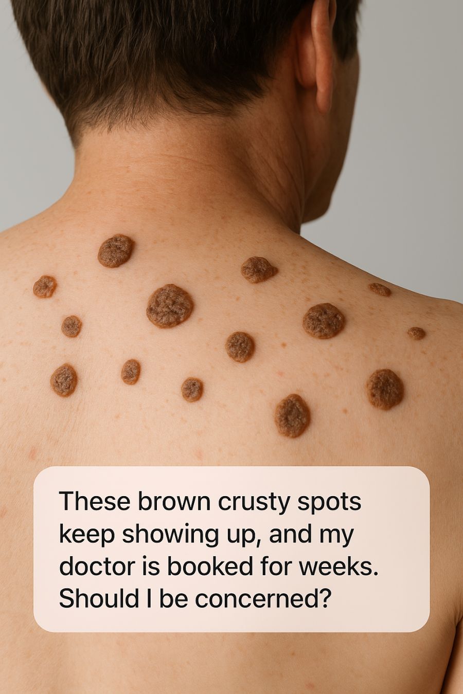
The brown, crusty spots you’re noticing are most often seborrheic keratoses, a very common, noncancerous skin growth that typically appears in adults over age 40. These growths range in color from light tan to dark brown and often have a rough, waxy, or wart-like texture. They may be only a few millimeters in size or grow larger than an inch.
Seborrheic keratoses are benign and usually don’t require treatment unless they become irritated or are cosmetically bothersome. Still, it’s important to monitor them for changes in size, shape, or color, which should be evaluated by a healthcare professional.
2. How to Tell Seborrheic Keratosis From Skin Cancer
Distinguishing seborrheic keratoses from skin cancer can be difficult without medical training. Seborrheic keratoses often have a waxy, “stuck-on” appearance, whereas melanoma may show irregular borders, uneven coloring, asymmetry, and changes over time.
Other skin cancers, such as basal cell or squamous cell carcinoma, may appear as non-healing sores, scaly patches, or areas that bleed easily. Any spot that bleeds, becomes painful, or changes rapidly should be examined by a medical professional.
3. The Classic Look: Color, Texture, and the “Stuck-On” Appearance
Seborrheic keratoses are known for their distinctive “pasted-on” look. They appear to sit on top of the skin rather than grow from within it, which helps differentiate them from other lesions.
Their color can range from pale tan to dark brown or nearly black, and their texture may be smooth, rough, or crumbly. While dermatologists can often identify them at a glance, professional evaluation is always best if there’s uncertainty.
4. Who Gets Seborrheic Keratoses—and Why They Multiply
Seborrheic keratoses are most common after age 40. Genetics play a strong role, meaning they often run in families. Sun exposure, hormonal changes, and skin friction may also contribute.
They tend to increase in number over time and may appear in clusters. Although a sudden increase can be alarming, it’s usually a normal part of aging. Still, noticeable changes should be monitored.
5. When Brown Spots Are a Red Flag
Most brown spots are harmless, but certain features should not be ignored. Seek medical evaluation if a spot bleeds, becomes painful, shows multiple colors, or changes rapidly in size or shape.
Another warning sign is the sudden appearance of many new lesions in a short period of time, which may indicate an underlying condition. When in doubt, it’s safest to consult a healthcare provider.
6. Can You Wait for Your Appointment—or Is It Urgent?
In most cases, seborrheic keratoses are not urgent and can safely wait until your scheduled dermatology visit. However, lesions that bleed, grow quickly, or cause significant discomfort should be evaluated sooner.
Those with a personal or family history of skin cancer should be especially cautious and seek earlier assessment if changes occur.
7. Why TikTok and DIY Removal Hacks Are Risky
Social media is full of do-it-yourself skin remedies, but attempting to remove skin lesions at home can be dangerous. DIY methods may cause infection, scarring, or delayed diagnosis of serious conditions like skin cancer.
Dermatologists use sterile tools and techniques to safely remove growths and confirm diagnoses—something that can’t be replicated at home.
8. How Dermatologists Diagnose These Spots
A dermatologist will perform a full skin examination, often using a dermatoscope, which magnifies and illuminates the skin to reveal detailed patterns.
If there’s any uncertainty, a biopsy may be performed. This involves removing a small sample of tissue for laboratory analysis and is typically quick and done with local anesthesia.
9. Treatment Options: From Removal to Watchful Waiting
Seborrheic keratoses usually don’t require treatment. When removal is desired, options include cryotherapy (freezing with liquid nitrogen), curettage (gentle scraping), or electrosurgery.
Your dermatologist will recommend the most appropriate method based on the lesion’s size, location, and your preferences.
10. Safe At-Home Skin Checks While You Wait
Perform regular self-exams using mirrors or assistance from someone you trust. Look for changes in size, shape, color, texture, or symptoms like bleeding or itching.
Keeping notes or photos of changes can be helpful for your appointment. Protect your skin with sunscreen of SPF 30 or higher while you wait.
11. How to Talk to Your Doctor So Your Concerns Are Heard
Be specific when discussing your spots—mention when they appeared, how they’ve changed, and whether there’s a family history of skin cancer. Photos showing changes over time can be very helpful.
Ask questions about possible diagnoses and treatment options, and don’t hesitate to seek a second opinion if you feel unsure.
12. Protecting Your Skin Going Forward
Long-term skin protection is essential. Use broad-spectrum sunscreen daily, wear protective clothing, and avoid peak sun hours when possible.
Continue monthly self-exams and schedule regular dermatology visits, especially if you have a history of skin issues. Early detection is one of the most effective tools for maintaining healthy skin.
